The narrow neck between two joints
Each vertebra has a heavy block at the front and a ring of bone behind it. Along each side of the ring sit two small facet joints, one facing up and one facing down. The stretch of bone between them is the pars interarticularis, usually just the pars, and the name is Latin for the part between the joints.
It is a narrow piece of bone with a demanding job. It is the strut that ties the block at the front to the joints at the back, and it takes strain whenever you lean back or twist, most of all in the lowest lumbar levels. On an angled X-ray the outline of a vertebra is said to look like a Scottie dog, with the pars as its neck, and a defect shows as a collar.
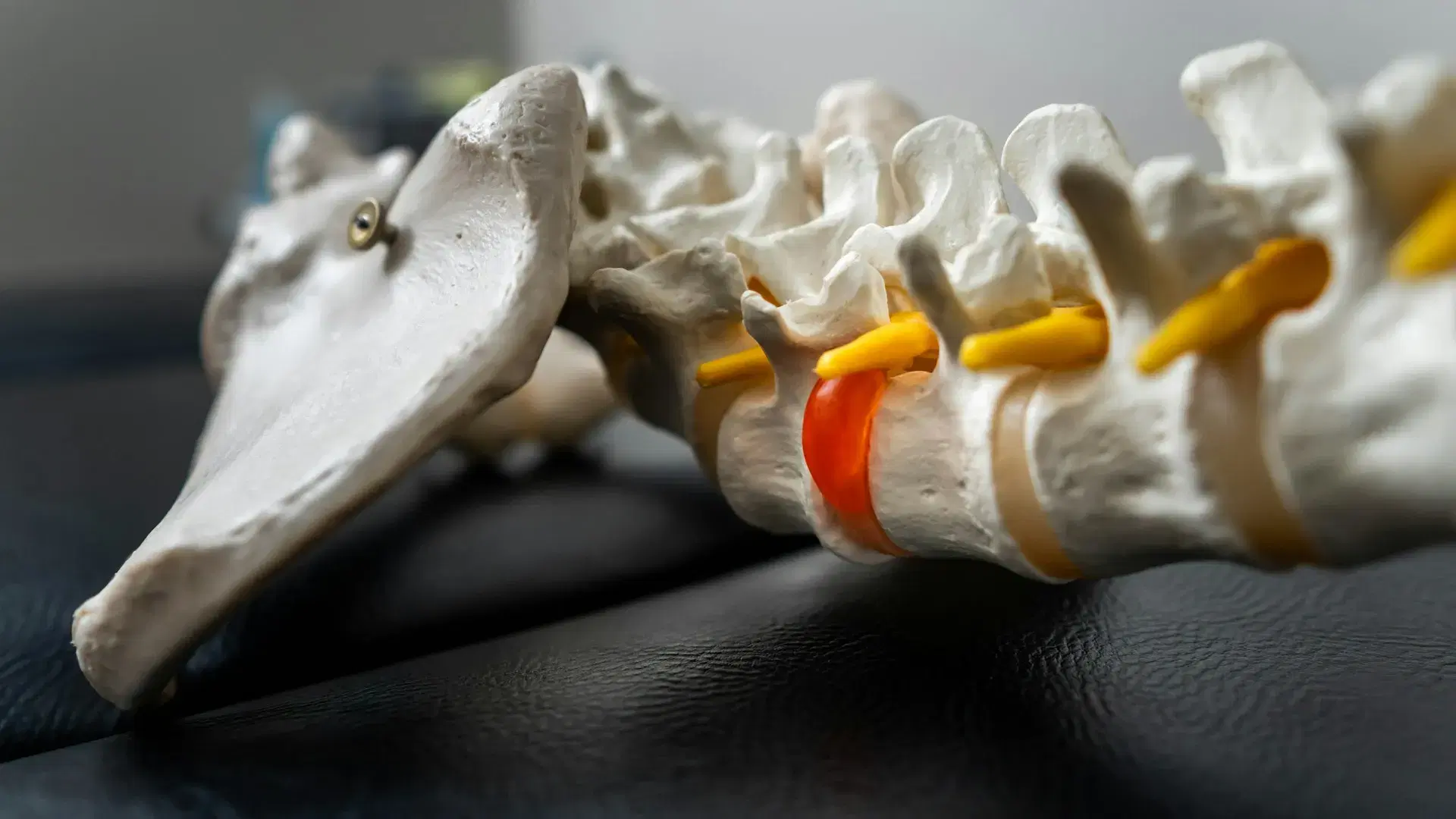
The lowest lumbar vertebrae, L4 and L5, above the sacrum, seen from the left side with the front of the body on the left. On L5, the bony ring behind the body has an upper facet and a lower facet, and the narrow bridge between them is the pars. That bridge is highlighted and drawn cut across by a small gap, so on this side the front of the vertebra and the back of the ring are no longer joined. The body has not slipped forward: it still sits where it should on the sacrum.
How a gap forms, and why it is usually old
A pars defect is thought to start as a stress injury. Repeated bending backwards and twisting load the pars until it cracks, as a paperclip bent back and forth finally gives. Many defects are believed to form earlier in life and never knit, which is why on an adult’s scan the edges are often smooth and rounded, with denser bone around them, the look of something long standing. A person can carry one for decades without knowing.
A recent one looks different. On an MRI, fluid in the bone around the pars can mark a stress reaction, and it tends to come with pain that began after a sudden rise in a sport that repeats arching and twisting, such as gymnastics or fast bowling. That is a question for your GP or a sports doctor.
Found by chance: reading the line on your report
A report line usually carries three facts: the level, which is most often L5 and next most often L4; whether the gap is on one side or both; and whether the bone in front has moved. “No listhesis” means nothing has slipped, which is worth looking for in any line about a defect.
Many are found by chance, on a scan ordered for leg pain or a disc, which is why a defect can appear on the report of someone whose pain is plainly elsewhere. A scan is arranged when clinically indicated. CT shows bone in the most detail, and MRI shows fluid in the bone and the nerves beside it. The Australian Commission on Safety and Quality in Health Care’s Low Back Pain Clinical Care Standard (2022) says what to expect from imaging, and its limited role in diagnosing low back pain, should be talked through with you. A defect turned up by a scan for something else shows why: the line on the report has to be read beside an examination of your back.
A defect and a slip are different findings
The pars is the strut that stops the front block sliding away from the back ring. With a gap on both sides, the block is free of the ring and can move forward, carrying the vertebra above with it. A slip that follows a gap is called isthmic spondylolisthesis, after the isthmus, the word for a narrow connecting strip, and like any slip it is graded by how many quarters of the bone below the upper vertebra has moved forward.
Many gaps never slip. A slip can also happen without any break in the bone, when wear loosens the joints and the whole vertebra drifts forward. So the two findings overlap without being the same, and either can be reported alone.
When it is the sore part, and when it is only in the report
A defect can be silent. When it is the sore part, the usual picture is an ache low in the back on one side or both, brought on by leaning back, standing a long time or loaded arching, and quieter when you bend forward. The hamstrings may feel tight. Leg symptoms are not typical unless a slip has narrowed an exit for a nerve.
A defect that has been there for years can sit in a back where the pain comes from somewhere else, a disc, a facet joint or a muscle, and the same picture can show both. The examination has to separate them, and the facet joints, which sit just either side of the pars, are the near neighbour to rule in or out.
How the report line is weighed against the back
The back is asked to lean backward, sometimes on one leg at a time, because that position loads the pars most. Pain provoked there on one side is a clue and not proof. The hamstrings, hips and leg nerves are examined too, since the same report may name a disc and a joint as well.
Where a defect fits, it shapes which hands-on techniques are suitable at that level and which are left out, and what is asked of the muscles that steady the trunk. A defect that is old and quiet is part of the picture of the back, not an instruction to stop moving.
Words about the bone bridge on a report
The words you are likely to meet on a report about this bone. Whoever ordered the scan is the one to read them against your history.
- Pars interarticularis, the pars
- The bridge of bone between the upper and lower facet joints of a vertebra. It is where the defect sits.
- Spondylolysis
- The formal word for a break or gap in the pars. A pars defect and spondylolysis are one finding with two names.
- Unilateral, bilateral
- One side or both. With a gap on both sides the front block is free of the back ring. With a gap on one side the other side still holds them together.
- Pars stress reaction
- Fluid in the bone at the pars on an MRI, before or without a full gap. It is the report’s word for a recent stress injury in the bone.
- Sclerosis
- Denser bone around the gap, which looks white on an X-ray or CT. It is often how an older defect looks.
- Listhesis
- A slip of one vertebra on the next. “No listhesis” means no slip was seen.
- Isthmic
- From isthmus, a narrow connecting strip. Isthmic spondylolisthesis is a slip that follows a gap in the pars.
When to get it looked at
Go to a hospital emergency department straight away if you have numbness or altered feeling around the saddle area, meaning the groin, the inner thighs, the buttocks or the genitals; a loss of normal sensation in the genitals, including during sex; trouble starting to pass urine, or any new change in your bladder or bowel control; weakness in both legs, particularly if it is getting worse; new or worsening pain running down both legs; weakness in one leg that is severe or getting worse, or a foot that has started to catch or drag when you walk; or numbness in a leg that is spreading or getting worse quickly.
Go to a hospital emergency department straight away for back or neck pain after a serious fall or a high-impact accident, such as a car accident; back or neck pain with a fever or feeling generally unwell; or new back or neck pain if your immune system is weakened, for instance by long-term steroids or chemotherapy, if you inject drugs, or if you have recently had a spinal injection, spinal surgery or a dental procedure.
Go to a hospital emergency department the same day if you have new clumsiness in both hands, or new unsteadiness on your feet.
See your GP promptly, before booking here, if you have a history of cancer, unexplained weight loss, pain at night that will not settle in any position, known osteoporosis with new severe pain, numbness that is spreading or getting worse slowly, or new, unexplained back pain as an older adult.
If a pars defect is on your report and the ache is one-sided, returns with leaning back or standing, or comes with tight hamstrings, ask for the report to be read against an examination of your back. If the ache began recently, after a sudden rise in loading, see your GP first, because that raises the question of whether the bone needs imaging. Earlier reports are worth having to hand as well.