

A spine MRI report has three working parts: the technique, which says what was scanned and how; the findings, which list what the radiologist saw at each level; and the impression, which sums up what matters. The findings list is written to be complete, so it reads heavier than it feels, and it needs matching to your examination.
The top of the report: what was scanned and why
Before any finding there is a block that is easy to skip and worth a minute. It names the scan, for example MRI lumbar spine, along with the date, the person who asked for it and a line of clinical details: the reason the scan was ordered, in their words. If the clinical details say “right leg pain” and the pain that bothers you now is on the left, the report was written to answer a different question from yours.
Next comes the technique. It lists the sequences, such as T1 and T2 weighted images in the sagittal plane (the side view) and the axial plane (slices across the spine), and says whether a contrast dye was used. Most of that is written for other doctors. What is worth noticing is whether an earlier scan was compared, because a line such as “no significant change since the previous study” is a finding of its own, and an old report read on its own can mislead.
The findings, level by level
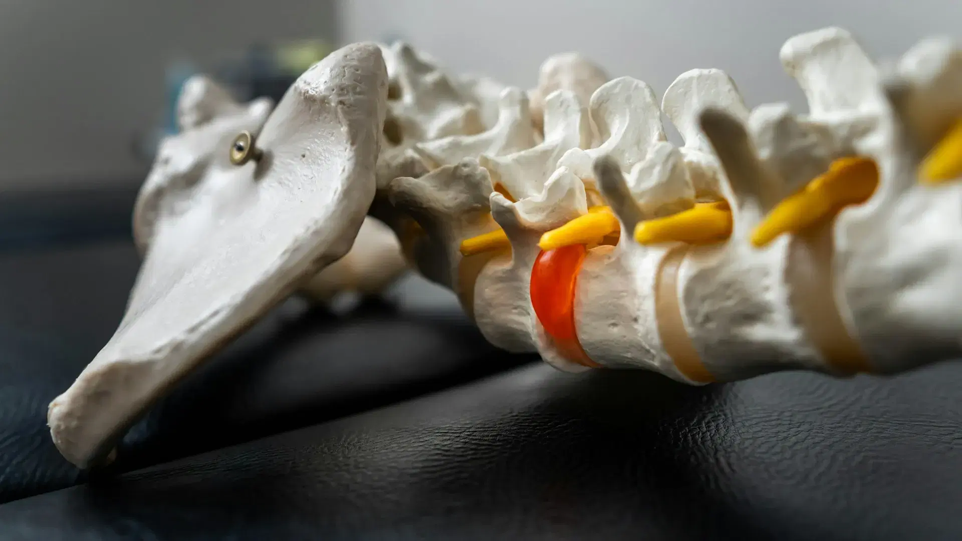
Findings are organised by level, and levels are named by the bones on either side of a disc. L4/5 is the disc between the fourth and fifth vertebrae of the lumbar spine, and L5/S1 sits between the last lumbar vertebra and the sacrum. In the cervical spine the same pattern gives C5/6, and in the thoracic spine T7/8. The same level may be written L4-5 or L4–L5. The vertebrae are counted from the top of each region: seven in the neck, twelve in the mid back and five in the lower back.
Each level gets a short paragraph that covers the same handful of things, so it helps to know what they are:
- Alignment. Whether the vertebrae line up in a smooth curve. Spondylolisthesis is the word for one vertebra sitting forward of the one below it. On a neck report, loss of cervical lordosis means the usual curve looks flatter than expected on that picture. On a thoracic report, kyphosis is the name of the outward curve every upper back has, so read the words around it.
- The bones and their marrow. The height, shape and signal of each vertebra. Modic changes are signal changes in the marrow beside a disc, described by type. A small pocket where disc material has pushed into the bone is a Schmorl’s node, and a gap in the narrow bridge of bone at the back of a vertebra is a pars defect, written as spondylolysis.
- The discs. Their height and how much water they appear to hold. A disc that holds less water is described as desiccated, a finding written as disc desiccation, and a lower, drier disc is often written up as degenerative disc disease, a name that sounds like a diagnosis and describes changes that become more common with age.
- The disc wall and its edge. A small split in the outer wall of a disc is called an annular tear or fissure. Disc material that reaches past the edge is named by its shape: a bulge is broad and shallow, a disc protrusion is more focused, and an extrusion has pushed further out.
- The canal and the side openings. The central canal carries the nerves, and the foramina are the openings at each level where a nerve root leaves. Narrowing of the canal is called spinal stenosis, and narrowing of an opening is foraminal stenosis.
- The joints and the bone edges. The small paired joints at the back of each level are the facet joints, and wear at those joints is written as facet arthropathy. Bony spurs are called osteophytes, and spondylosis is the report’s general word for wear-related change, written as cervical spondylosis on a neck report. If the scan takes in the pelvis, the sacroiliac joint may get a line too. Sacroiliitis means that joint looks inflamed, and that word is one for your GP.
- The nerve roots. Whether a root is touched, shifted or squeezed, which a report may call nerve root compression. A scan can show a root being touched. Whether the root is struggling, which is what radiculopathy means, is judged from your symptoms and the examination. In the lower back the structures in the canal are nerve roots, because the spinal cord ends higher up, near the top of the lumbar spine. They sit inside a fluid-filled sleeve, the thecal sac, so “indenting the thecal sac” describes something touching that sleeve.
Reports also grade how far a change goes, with words such as mild or severe. Those describe the size of the change, not how much it hurts. A severe-sounding word on a quiet level can sit beside a sore level the report calls mild, and the two do not have to match.
Why the findings list reads worse than it feels
A radiologist’s job is to report everything visible, so the findings section is written to be complete and not to rank importance. A change that has been there for years and a change that matters this month are set out in the same flat voice. Long lists of long words are the result.
Many of the changes a report lists, such as disc bulges, discs that hold less water and wear at the joints, also show up on the scans of people who have no back pain, and they become more common with age. It is one reason to ask whether you need a scan first. The practical consequence is that a finding means something only when it matches your story and your examination. Where it matches, it can be part of the explanation. Where it does not, it describes your spine and not your pain.
The vocabulary adds to the weight. Degeneration, bulge and tear are the radiologist’s names for appearances, drawn from a standard set, and they carry more alarm in everyday English than they do on the report. Often a disc described as degenerate is showing its age, the way a fifty-year-old hand looks older than a twenty-year-old one.
The Australian standard is just as cautious. The Australian Commission on Safety and Quality in Health Care’s Low Back Pain Clinical Care Standard (2022) says, in its third quality statement, that what to expect from imaging, and its limited role in diagnosing low back pain, should be talked through with you. The Royal Australian College of General Practitioners’ First do no harm: A guide to choosing wisely in general practice. For GPs: Imaging in adults with acute low back pain (2022) adds that for acute low back pain, imaging is recommended only after a careful clinical assessment raises a high suspicion of a serious cause. The clinic requests imaging when clinically indicated, not as a matter of routine.
Not every line is background. If a report mentions a fracture, an infection, a mass or lesion, or pressure on the spinal cord or the cauda equina (the bundle of nerve roots at the bottom of the canal), take it to whoever ordered the scan promptly, before booking here. If you also have symptoms that need an ambulance, an emergency department or your GP, those come first.
The impression, and what to do with the report
The impression, sometimes headed conclusion, comment or summary, is the radiologist’s own shortlist. It is written for the doctor who asked for the scan and picks out the findings that bear on the clinical question. It is reasonable to read it first, so long as you go back to the findings afterwards with the level names in mind.
Four questions are worth putting to whoever ordered the scan, or at an assessment:
- Which finding, if any, sits at the level that matches where you hurt?
- Which findings are changes that come with age, and does any of them call for action?
- Does anything in the report change what happens next?
- Does the report mention anything outside the spine that needs following up?
Bring the written report to an assessment, with the images if you have them, and say when the scan was done. It is read alongside the examination and not in place of it. A report describes a spine on the day it was scanned, so what you feel now and what the examination shows now carry more weight than a line from an old one.
A report is only a description. Whoever ordered the scan is still the one who follows it up, and no report can say by itself what is causing your pain or how you move.
What the guidelines say
- Australian Commission on Safety and Quality in Health Care, Low Back Pain Clinical Care Standard, 2022. https://www.safetyandquality.gov.au/standards/clinical-care-standards/low-back-pain-clinical-care-standard
- Royal Australian College of General Practitioners, First do no harm: A guide to choosing wisely in general practice. For GPs: Imaging in adults with acute low back pain, 2022. https://www.racgp.org.au/clinical-resources/clinical-guidelines/key-racgp-guidelines/view-all-racgp-guidelines/first-do-no-harm/gp-resources/imaging-in-adults-with-acute-low-back-pain
When to get it looked at
Call an ambulance if there is any confusion, sudden weakness or numbness in the face, arm or leg on one side, facial drooping, slurred speech, sudden trouble speaking, seeing or understanding, loss of vision, a seizure, or a sudden loss of balance or coordination.
Call an ambulance or go to a hospital emergency department straight away if a headache comes on suddenly and severely (the worst you have ever had), if a headache arrives with fever, a stiff neck or a rash, or if a headache follows a knock to the head.
Go to a hospital emergency department straight away if you have numbness or altered feeling around the saddle area, meaning the groin, the inner thighs, the buttocks or the genitals; a loss of normal sensation in the genitals, including during sex; trouble starting to pass urine, or any new change in your bladder or bowel control; weakness in both legs, particularly if it is getting worse; new or worsening pain running down both legs; weakness in one leg that is severe or getting worse, or a foot that has started to catch or drag when you walk; or numbness in a leg that is spreading or getting worse quickly.
Go to a hospital emergency department straight away for back or neck pain after a serious fall or a high-impact accident, such as a car accident; back or neck pain with a fever or feeling generally unwell; or new back or neck pain if your immune system is weakened, for instance by long-term steroids or chemotherapy, if you inject drugs, or if you have recently had a spinal injection, spinal surgery or a dental procedure.
Go to a hospital emergency department straight away if you have weakness in one arm that is severe or getting worse, or numbness in an arm that is spreading or getting worse quickly.
Go to a hospital emergency department the same day if you have new clumsiness in both hands, or new unsteadiness on your feet.
See your GP promptly, before booking here, if you have a history of cancer, unexplained weight loss, pain at night that will not settle in any position, known osteoporosis with new severe pain, numbness that is spreading or getting worse slowly, new, unexplained back pain as an older adult, or pins and needles, numbness or weakness in an arm or hand.
If any warning sign applies to you, the report can wait: the hospital or your GP comes first. A scan result that has not been explained to you belongs with whoever ordered it. Beyond that, a report is worth bringing to an assessment when the pain it describes has lasted or keeps returning, and you want the findings matched to how you move and what you do.