

Bone adds an edge where a joint takes load
A joint surface is covered in cartilage, and over the years that cartilage thins. Where it does, the bone at the margin lays down more bone, which widens the area that carries the load. The result is a lip, a shelf or a beak at the edge of the joint. Spur is a harsh word for it. On an image it is more often a smooth rim than a thorn, and it forms over a long time rather than in a day.
Osteophyte means bone growth, and osteophytosis is the report’s word for having them. Growth is a worrying word, but this one means bone remodelling at a worn edge, not a tumour. You may also meet spurring, lipping or beaking, which are the same thing in other words.
Where they form on the spine
- The edges of the vertebral bodies, at the front, sides and back, where each bone meets a disc. A run of them across several levels is part of what reports call spondylosis.
- The facet joints at the back, where an enlarged joint and its spurs tend to go together, and the wear in the joint itself is what a report may call facet arthropathy.
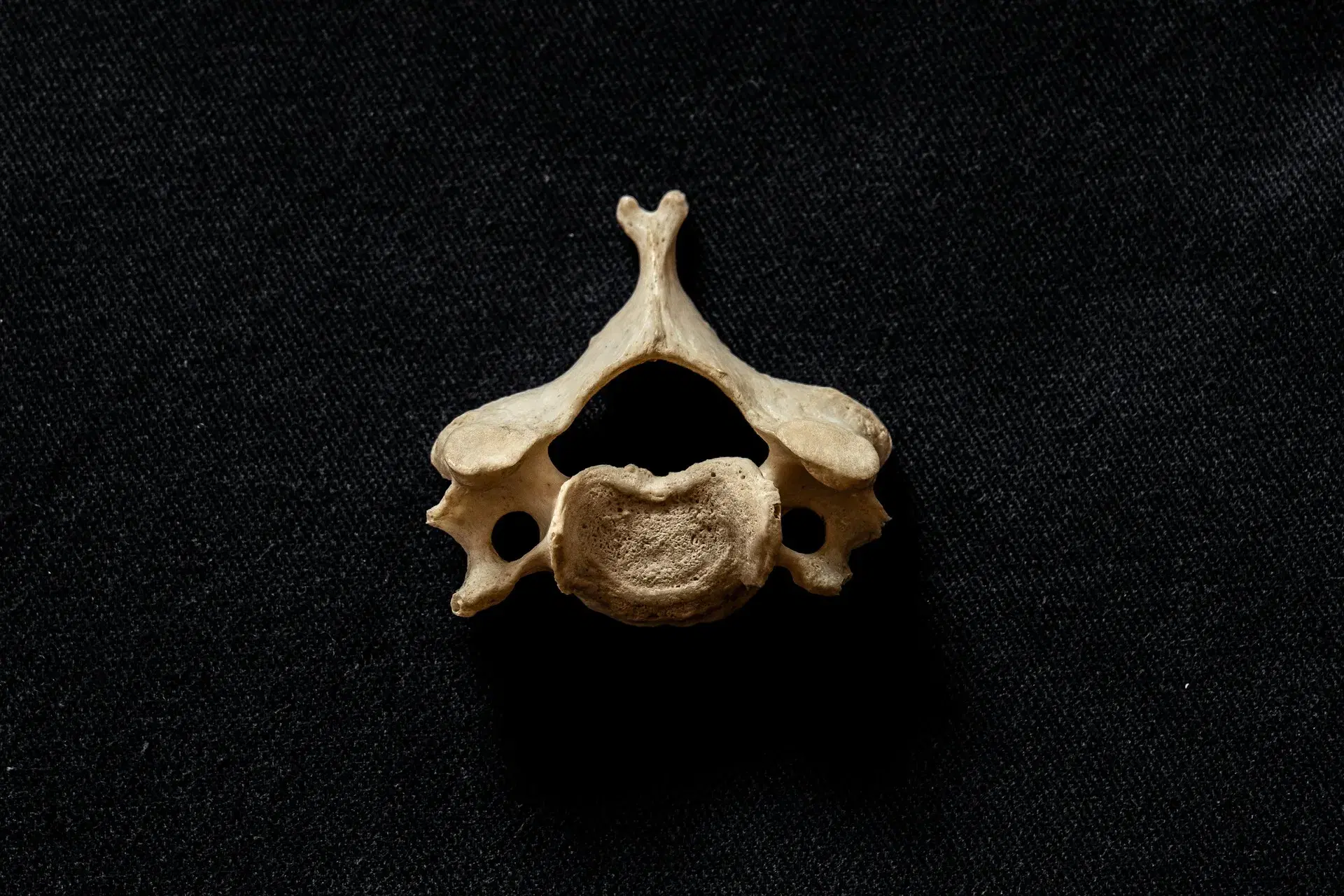
- The small side joints of the neck, which sit just in front of the exit for each nerve root and can narrow it.
- The rim of a disc space that has lost height, because bone responds where the load has moved.
Two lumbar vertebrae, L4 above L5, seen from the left side with the front of the body on the left, and the disc between their bodies. Small beaks of new bone, highlighted, grow from the rim of each body beside the disc: two at the front and two at the back. The back ones lean toward the opening behind the disc, where a nerve root leaves the canal; the root itself is left out so the opening can be seen.
Why a spur on its own says little
A report is a list of what is visible, and a spur is easy to see. That makes it a prominent line on a report and a weak suspect by itself. A spur can sit at a joint that has never given trouble, and a joint can hurt without one. Whether the bone is the source of anything is a separate question that a picture cannot answer.
The same goes for how a spur is described. Small, mild or moderate describe size on the image, not how a person feels, and a larger spur is not automatically a sorer one. The Australian Commission on Safety and Quality in Health Care’s Low Back Pain Clinical Care Standard (2022) says what to expect from imaging, and its limited role in diagnosing low back pain, should be talked through with you, and a spur in the lower back is a fair example of why. The clinic requests imaging when clinically indicated, not to go looking for spurs. Every other size word on a report works the same way. If you have one in front of you, it helps to know how to read a spine MRI report.
Position is what makes one matter
What counts is what the spur sits beside. A spur that points away from the nerves is usually just a feature of the bone. One that points toward a nerve has more room to be relevant, and a report may say where it points.
- Into the central canal. It adds to a narrowing of the canal that holds the nerves, which a report calls spinal stenosis.
- Into a nerve exit. It can narrow the side opening a root passes through, which is what foraminal stenosis describes.
- Onto a facet joint. It widens the joint, and that joint may be stiffer or more tender when pressed.
Even then the spur is only part of the picture. A disc or an inflamed joint can irritate the same root, and the spur may be a bystander. The examination links a finding to a nerve: which fingers or toes are involved, which muscle is weak, what the reflexes do. A spur in the neck beside an arm with pins and needles is one way a pinched nerve in the neck comes about, and that arm goes to your GP first. One more position counts, at the front of the neck: a large spur there can occasionally press on the gullet, so new trouble swallowing is one for your GP.
Reading a spur against how you move
If a report names a spur, bring it. The assessment starts with what you feel and when, then looks at how the region moves, which muscles serve it, and the strength, reflexes and sensation of the nerves nearby. A spur that fits those findings is part of the explanation. A spur that does not stays on the report as a description, and the examination carries on looking for the source.
Words a report uses for bone growth
They describe the picture. Whoever ordered the image is the one to read them against your history.
- Osteophyte, spur
- A bony outgrowth at the margin of a joint. Both words describe shape and place on the image.
- Osteophytosis
- The state of having osteophytes. A report may use it for one joint, one level or a whole region.
- Marginal osteophyte
- A spur at the rim of a vertebral body, where the bone meets the disc.
- Facet hypertrophy
- Enlargement of the small joints at the back of the spine, often with spurs at their edges.
- Uncovertebral hypertrophy
- Enlargement of the small joints at the sides of the neck vertebrae, which lie beside the exit for the nerve root.
- Foraminal encroachment
- A spur or other structure reaching into the exit opening for a nerve. It describes position, not pain.
- Bridging osteophytes
- Spurs from two neighbouring vertebrae that meet across the disc space. It names a stage of change at that level.
When to get it looked at
Call an ambulance if there is any confusion, sudden weakness or numbness in the face, arm or leg on one side, facial drooping, slurred speech, sudden trouble speaking, seeing or understanding, loss of vision, a seizure, or a sudden loss of balance or coordination.
Call an ambulance or go to a hospital emergency department straight away if a headache comes on suddenly and severely (the worst you have ever had), if a headache arrives with fever, a stiff neck or a rash, or if a headache follows a knock to the head.
Go to a hospital emergency department straight away if you have numbness or altered feeling around the saddle area, meaning the groin, the inner thighs, the buttocks or the genitals; a loss of normal sensation in the genitals, including during sex; trouble starting to pass urine, or any new change in your bladder or bowel control; weakness in both legs, particularly if it is getting worse; new or worsening pain running down both legs; weakness in one leg that is severe or getting worse, or a foot that has started to catch or drag when you walk; or numbness in a leg that is spreading or getting worse quickly.
Go to a hospital emergency department straight away for back or neck pain after a serious fall or a high-impact accident, such as a car accident; back or neck pain with a fever or feeling generally unwell; or new back or neck pain if your immune system is weakened, for instance by long-term steroids or chemotherapy, if you inject drugs, or if you have recently had a spinal injection, spinal surgery or a dental procedure.
Go to a hospital emergency department straight away if you have weakness in one arm that is severe or getting worse, or numbness in an arm that is spreading or getting worse quickly.
Go to a hospital emergency department the same day if you have new clumsiness in both hands, or new unsteadiness on your feet.
See your GP promptly, before booking here, if you have a history of cancer, unexplained weight loss, pain at night that will not settle in any position, known osteoporosis with new severe pain, numbness that is spreading or getting worse slowly, new, unexplained back pain as an older adult, or pins and needles, numbness or weakness in an arm or hand.
A spur that sits quietly on a report is a note on a picture. One that keeps company with pain or stiffness at the same level, coming back again and again, or with leg symptoms you can describe, is the kind an examination can weigh. If the spur is in the neck and an arm or hand has pins and needles, numbness or weakness, your GP sees that first. Bring the report, so that its wording can be read against how you move and not taken as the explanation by itself.